Carnegie Microscopy Suite
The microscopy suite is a shared campus facility used primarily by the departments of Biology, Earth and Climate Sciences, Neuroscience, Physics, and Chemistry.
Microscopy Lab User Policy ’20-’21 | Training | Equipment |
Connect2 microscope reservations
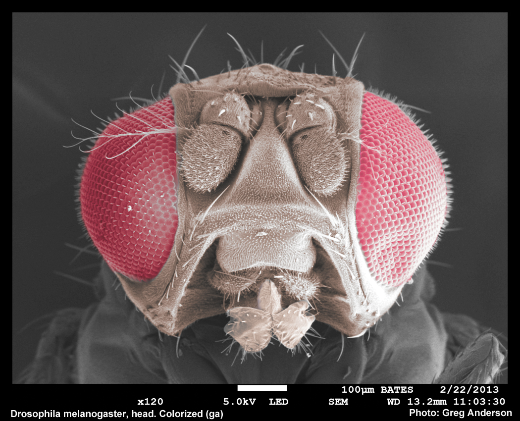
Wildtype Drosophila melanogaster head. Colorized post-acquisition using Adobe Photoshop.
During fall 2012 the lab acquired a new FE-SEM/EDS system funded in large part by a Sherman-Fairchild Foundation grant to Bates College. The college has purchased a JEOL JSM-7100 FLV variable vacuum field emission SEM and a Thermo/Noran System 7 EDS system.
In the fall of 2014, a new Leica SP8 Laser Scanning Confocal Microscope came on line which was funded by a NSF-MRI grant to Larissa Williams, Matt Cote, Nancy Kleckner, and Travis Gould.
As part of a STEM facilities upgrade, in summer 2018, the light and fluorescence microscopes from the Imaging Center were moved into the suite to create a true microscopy center.
We look forward to training our students on these up-to-date technologies and welcome the expanded opportunities for innovative teaching and research that the new instruments will bring!
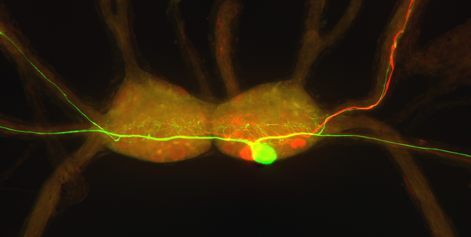
Confocal image of buccal ganglia of the tropical pond snail, Biomphalaria glabrata.
SEM/EDS Training Contact:
Greg Anderson, AI, EM lab tech
(207) 786-6110
ganderso-at-bates.edu
Confocal Microscope Training Contact:
Dr. Travis Gould, Facility Director
Physics Department
tgould-at-bates.edu